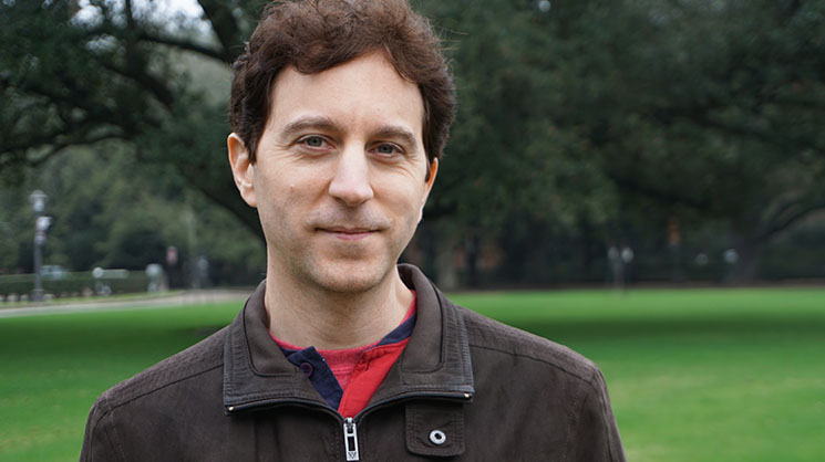
Didier Devaurs, a postdoctoral research associate at Rice University, has a passion for interdisciplinary research.
Bioinformatics linked his computational experience, applying geometrical techniques to modeling, with his interest in chemistry and biology. Devaurs was born in Aurillac, France but his work experience has taken place in Spain, Austria and Canada.
“There are not that many places with this particular combination [robotics techniques applied to biology]. To be honest, Lydia Kavraki was the researcher at the top of my list, and I was so happy when she offered me a place here at Rice. I didn’t realize at the time how much Rice’s location across the street from the Texas Medical Center would improve my opportunities to connect with medical researchers,” he said.
The problem Devaurs and his colleagues are tackling involves mapping the rapid motions of molecules. Too tiny to track with a microscope, proteins can be modeled with techniques perfected in robotics.
“The idea itself is not new,” Devaurs said. “Comparing the rotation and folding or unfolding behaviors of proteins to robotic movement was suggested about 20 years ago. I’m just pushing the method.”
Like robotic or human arms, proteins can move in certain constrained ways. Both physical systems and proteins have different degrees of freedom; they can rotate in some ways, but not others. To map the motions for a robotic arm, researchers first consider how it can interact with its environment. To model protein behaviors, scientists must also consider how it interacts with other molecules in its environment.
“So the common points in both systems are to plan and then direct. What is the best way to move to meet the goal, and then how do we execute those movements? Our goal for the protein modeling is to provide a way for biomedical researchers to better understand how molecules interact with each other. The scale is way too small to view through a microscope, so the only recourse is to simulate the behavior in the most realistic way possible,” Devaurs said.
One of the experimental methods of exploring molecule three-dimensional structure is with X-ray crystallography. The process is expensive and produces a single image of a molecule in a frozen state. To project how the molecule arrived at that state and where it will go next is like trying to imagine how an ice skater completes a double axel by using a single photo taken during the leap.
Devaurs said the method he uses helps to explain and simulate molecule change.
“Molecules are always moving and changing their shape. Their shape is linked to their function, like for an enzyme chopping another molecule. These still images are high definition, but do not capture the dynamic life of the molecules. So what we bring to the picture is a possible explanation and simulation about how the molecules change shape and interact,” he said.
Devaurs continued, “Rice was definitely a good place for me to do the interdisciplinary work I was interested in from the beginning because of the Medical Center’s presence. The geographic proximity helps making these connections happen especially in my area, bioinformatics.”
